


Recombinant anti-PD-L1 antibodies (D265A) - InvivoFit™
| Product | Unit size | Cat. code | Docs. | Qty. | Price | |
|---|---|---|---|---|---|---|
|
Anti-mPD-L1-mIgG1e3 (10F.9G2) Mouse PD-L1 (10F.9G2) antibody - Mouse IgG1, effectorless - InvivoFit™ |
Show product |
1 mg 10 mg 50 mg |
mpdl1c2-mab15-1
|
|
||
|
Anti-PD-L1-mIgG1e3 (Atezo) Mouse/human PD-L1 (Atezolizumab) antibody - Mouse IgG1, effectorless - InvivoFit™ |
Show product |
1 mg 10 mg 50 mg |
pdl1-mab15-1
|
|

InvivoGen’s engineered Anti-PD-L1-mIgG1e3 InvivoFit™ antibodies
 InvivoGen also offers:
Recombinant anti-PD-L1 antibodies for in vivo use
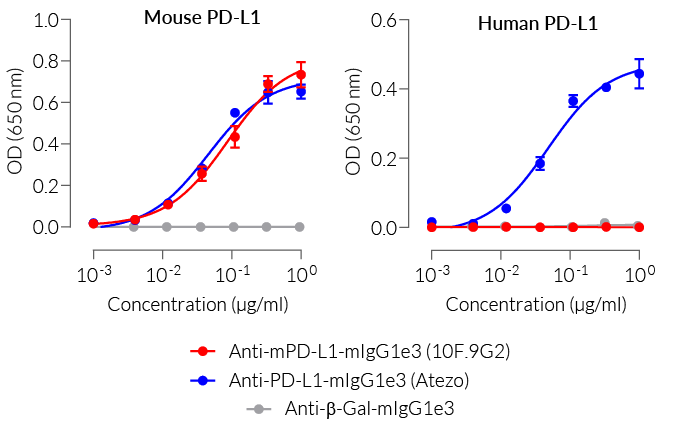
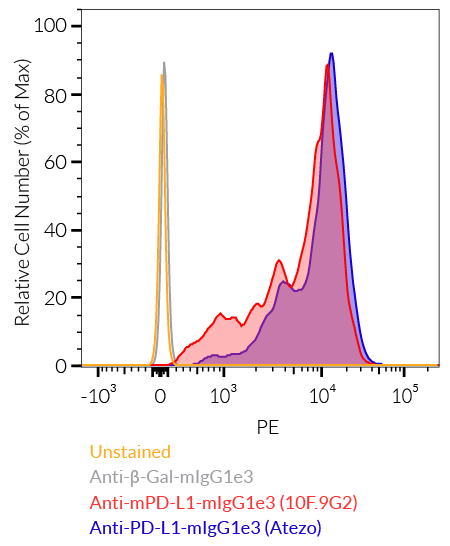
InvivoGen offers two murinized monoclonal antibodies (mAb) targeting PD-L1 (programmed cell death ligand 1), and therefore blocking the interaction with its receptor PD-1:
-
Anti-PD-L1-mIgG1e3 (10F.9G2) InvivoFit™ features the variable region of the rat-derived clone 10F.9G2, targeting mouse PD-L1 [1].
- Anti-PD-L1-mIgG1e3 (Atezo) InvivoFit™ features the variable region of atezolizumab, recognizing human and mouse PD-L1 [2].
The original anti-mPD-L1-rIgG2b (clone 10F.9G2) is the most commonly used PD-L1-targeting mAb in mouse in vivo studies. Atezolizumab (anti-PD-L1-hIgG1, also known as MPDL3280A) is a fully humanized therapeutic mAb, approved by the FDA for the treatment of various types of cancer [3]. Interestingly, it binds both human and mouse PD-L1. Upon repeated injections into mice, these mAbs can lead to immunogenicity and risks of fatal hypersensitivity reaction, since they are humanized and rat-derived, respectively [4].
To overcome these issues, InvivoGen replaced the humanized (Atezo) and rat-derived (10F.9G2) constant regions with a murine constant region, making the mAbs ~65% of murine origin. Both mAbs now harbor an engineered murine IgG1 constant region (mIgG1e3). Within that region, a point mutation was introduced changing the aspartic (D) acid at position 265 to an alanine (A) (D265A). This leads to a complete loss of unwanted Fc-associated effector functions [5]. These alterations increase antibody performance and overcome immunogenic events, especially in vivo [6].
Both antibodies are produced in Chinese hamster ovary (CHO) cells and purified by affinity chromatography with protein A. Its binding is validated by flow cytometry and ELISA. InvivoGen's anti-PD-L1 mAbs are provided in an InvivoFit™ grade, a high-quality standard specifically adapted to in vivo studies.
Key features of Anti-PD-L1-mIgG1e3 InvivoFit™ antibodies
- Derived from well-referenced clones: 10F.9G2 or Atezolizumab (aka MPDL3280A)
- Feature mouse IgG1e3 isotype (constant region)
- mIgG1e3 (IgG1 with a D265A point mutation) is effectorless
- Filter-sterilized (0.2 µm), endotoxin level: 1 EU/mg
- Suitable for parenteral delivery in mice (azide-free)
- Low aggregation < 5%
-
Produced in animal-free facilities and defined media
Programmed cell death ligand 1 (PD-L1; also called B7-H1 or CD274) binds to programmed cell death protein 1 (PD-1) on T cells and contributes to T cell exhaustion during chronic infections. Moreover, the engagement of PD-1 on T cells and PD-L1 on malignant cells is associated with the immune escape of tumors. Clinical trials have highlighted the anti-tumor efficacy of blockades targeting the PD-1/PD-L1 pathway [7].
References:
1. Eppihimer MJ, et al. 2002. Expression and regulation of the PD-L1 immunoinhibitory molecule on microvascular endothelial cells. Microcirculation.9(2):133-45.
2. Lesniak W.G. et al., 2016. PD-L1 Detection in Tumors Using [(64)Cu]Atezolizumab with PET. Bioconjug Chem. 27(9):2103-10.
3. Emens LA, et al., 2019. Long-term Clinical Outcomes and Biomarker Analyses of Atezolizumab Therapy for Patients With Metastatic Triple-Negative Breast Cancer: A Phase 1 Study. JAMA Oncol. 5(1):74-82.
4. Mall C. et al., 2016. Repeated PD-1/PD-L1 monoclonal antibody administration induces fatal xenogenic hypersensitivity reactions in a murine model of breast cancer. Onco Immunol. 5(2):e1075114.
5. Baudino L. et al., 2008. Crucial role of aspartic acid at position 265 in the CH2 domain for murine IgG2a and IgG2b Fc-associated effector functions. J. Immunol. 181(9):6664-9.
6. Brüggemann M. et al., 1989. The immunogenicity of chimeric antibodies. J. Exp. Med. 170:2153-2157.
7. McDermott D. & Atkins M. 2013. PD-1 as a potential target in cancer therapy. Cancer Med. 2(5): 662–673.
Figures


Specifications
Specificity: Targets cells expressing murine or human PD-L1
Formulation: Lyophilized from 0.2 μm filtered solution in 150 mM sodium chloride, 20 mM sodium phosphate buffer with 5% saccharose
Clonality: Monoclonal antibodies
Isotype: Murine IgG1e3 (D265A mutation; no effector function), kappa
Control: mIgG1e3 InvivoFit™ isotype control
Source: CHO cells
Purity: Purified by affinity chromatography with protein A
Tested Applications: Flow cytometry and ELISA
Quality control:
- Binding confirmed by flow cytometry
- The complete sequence of these antibodies has been verified
- < 5% aggregates (confirmed by size exclusion chromatography)
- Endotoxin level <1 EU/mg (determined by the LAL assay)
Contents
Please note: each mAb is sold separately.
All products are filter-sterilized (0.2 µm), endotoxin-free, azide-free, and lyophilized.
Anti-PD-mL1-mIgG1e3 (10F.9G2) InvivoFit™
- mpdl1c2-mab15-1 : 1 mg
- mpdl1c2-mab15-10 : 10 mg
- mpdl1c2-mab15-50 : 50 mg (5 x 10 mg)
Anti-PD-L1-mIgG1e3 (Atezo) InvivoFit™
- pdl1-mab15-1 : 1 mg
- pdl1-mab15-10 : 10 mg
- pdl1-mab15-50 : 50 mg (5 x 10 mg)
![]() The product is shipped at room temperature.
The product is shipped at room temperature.
![]() Store lyophilized antibody at -20 °C.
Store lyophilized antibody at -20 °C.
![]() Lyophilized product is stable for at least 1 year.
Lyophilized product is stable for at least 1 year.
![]() Avoid repeated freeze-thaw cycles.
Avoid repeated freeze-thaw cycles.
InvivoFit™
InvivoFit™ is a high-quality standard specifically adapted for in vivo studies. InvivoFit™ products are filter-sterilized (0.2 µm) and filled under strict aseptic conditions in a clean room. The level of bacterial contaminants (endotoxins and lipoproteins) in each lot is verified using a LAL assay and a TLR2 and TLR4 reporter assay.
Back to the topDetails
Programmed cell death ligand 1 (PD-L1), also known as cluster of differentiation 274 (CD274) or B7 homolog 1 (B7-H1) is a transmembrane protein that can be constitutively expressed or induced in myeloid, lymphoid, and normal epithelial cells, as well as in cancer [1, 2]. PD-L1 is the principal ligand for programmed cell death protein 1 (PD-1) and under physiological conditions, this interaction is essential for immune tolerance to prevent excessive immune cell activity.
However, PD-L1 expression is an immune evasion mechanism exploited by various malignancies and is generally associated with poorer prognosis [3]. Specifically, overexpressed PD-L1 on tumor cells and tumor-infiltrating immune cells, such as macrophages, can bind to PD-1 on cytotoxic T cells, and ultimately inhibit the anti-tumor T cell response [2, 4]. Thus, due to PD-L1’s instrumental role in immune evasion by cancer cells, there are numerous inhibitors in development as promising immuno-oncology therapies. Notably, Atezolizumab (also known as MPDL3280A), a fully humanized IgG1 (N298A) mAb that blocks the interaction of PD-L1 with PD-1 and induces anti-tumor immune reactivation, has been approved by the FDA for combinational use in the treatment of lung and breast cancer [2, 5].
References
1. Juneja, V.R. et al. 2017. PD-L1 on tumor cells is sufficient for immune evasion in immunogenic tumors and inhibits CD8 T cell cytotoxicity. J Exp Med 214, 895-904.
2. Kythreotou, A. et al. 2018. PD-L1. J Clin Pathol 71, 189-194.
3. Sun, C. et al. 2018. Regulation and Function of the PD-L1 Checkpoint. Immunity 48, 434-452.
4. Lau, J. et al. 2017. Tumour and host cell PD-L1 is required to mediate suppression of anti-tumour immunity in mice. Nat Commun 8, 14572.
5. Heimes, A.S. & Schmidt, M. 2019. Atezolizumab for the treatment of triple-negative breast cancer. Expert Opin Investig Drugs 28, 1-5.