


Recombinant anti-mouse CD3 antibody
| Product | Unit size | Cat. code | Docs. | Qty. | Price | |
|---|---|---|---|---|---|---|
|
Anti-mCD3-mIgG2a InvivoFit™ 145-2C11-derived mouse monoclonal antibody against murine CD3 |
Show product |
1 mg 10 mg |
mcd3-mab10-1
|
|
Recombinant murinized CD3ε antibody for in vivo use
Anti-mCD3-mIgG2a InvivoFit™ is an anti-mCD3 monoclonal antibody featuring the variable region of the previously described 145-2C11 clone [1] and a murinized IgG2a constant region.

InvivoGen’s engineered Anti-mCD3-mIgG2a InvivoFit™ antibody
CD3 is a multimeric protein complex consisting of four polypeptides (CD3ε, CD3γ, CD3δ, and CD3ζ) that assemble as three dimers (εγ, εδ, and ζζ) [2]. The CD3 complex is a marker of T cells [2]. Upon antigen recognition, the TCR/CD3 complex on T cells triggers downstream intracellular signaling and participates in T cell activation [3].
The anti-mCD3ε 145-2C11 mAb is commonly used for cytometry detection and in vivo depletion of the T cell population [1]. Using recombinant technology, the original 145-2C11 Armenian Hamster IgG1 constant region has been replaced with a murine IgG2a format which mediates potent cytotoxic functions [4]. This murinization also allows for reduced immunogenicity of the administered mAb and risks of fatal hypersensitivity reactions [5-7].
Anti-mCD3-mIgG2a is provided in an InvivoFit™ grade, a high-quality standard specifically adapted to in vivo studies.
Key features OF ANTI-MCD3-MIGG2A INVIVOFIT™:
- Derives from the 145-2C11 clone, Armenian Hamster IgG1
- Features the mIgG2a isotype (constant region)
- Filter-sterilized (0.2 µm), endotoxin level < 1 EU/mg
- Suitable for parental delivery in mice (azide-free)
- Low aggregation < 5%
- Produced in animal-free facilities and defined media
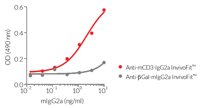
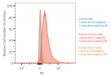
Anti-mCD3-mIgG2a InvivoFit™ is produced in Chinese hamster ovary (CHO) cells and purified by affinity chromatography with protein A. The specific binding of this mAb to mCD3 has been confirmed by FACS and ELISA (see Figures).
References:
1. Leo O., et al., 1986. Identification of a monoclonal antibody specific for a murine T3 polypeptide. PNAS. 84(5):1374-1378.
2. Chetty R. & Gatter K., 1994. CD3: structure, function, and role of immunostaining in clinical practice. J. Pathol. 173(4):303-307.
3. Smith-Garvin J.E. et al., 2009. T Cell Activation. Ann. Rev. Immunol. 27:591-619.
4. Nimmerjahn F. & Ravetch J.V., 2005. Divergent immunoglobulin g subclass activity through selective Fc receptor binding. Science. 310(5753):1510-2.
5. Mall C. et al., 2016. Repeated PD-1/PD-L1 monoclonal antibody administration induces fatal xenogenic hypersensitivity reactions in a murine model of breast cancer. Onco Immunol. 5(2):e1075114.
6. Murphy, J.T. et al., 2014. Anaphylaxis caused by repetitive doses of a GITR agonist monoclonal antibody in mice. Blood. 123(14):2172-2180.
7. Belmar N.A. et al., 2017. Murinization and H chain isotype matching of Anti-GITR antibody DTA-1 reduces immunogenicity-mediated anaphylaxis in C57BL/6 mice. J Immunol. 198:4502-4512.
Figures


Specifications
Specificity: Targets cells expressing murine CD3ε
Formulation: Lyophilized from 0.2 μm filtered solution in 150 mM sodium chloride, 20 mM sodium phosphate buffer with 5% saccharose
Clonality: Monoclonal antibody
Isotype: Murine IgG2a, kappa
Control: mIgG2a InvivoFit™ isotype control
Source: CHO cells
Purity: Purified by affinity chromatography with protein A
Applications: Flow cytometry; in vivo depletion; ELISA
Quality control:
- The binding of Anti-mCD3-mIgG2a InvivoFit™ to mCD3ε has been confirmed using Flow cytometry
- Antibody binding has been validated by ELISA using a coated mouse CD3 protein
- The complete sequence of the antibody construct has been verified
- < 5% aggregates (confirmed by size exclusion chromatography)
- Endotoxin level <1 EU/mg (determined by the LAL assay)
Contents
Anti-mCD3-mIgG2a InvivoFit™ is filter-sterilized (0.2 µm), endotoxin-free, azide-free, and lyophilized.
This product is available in two pack sizes:
- mcd3-mab10-1: 1 mg
- mcd3-mab10-10: 10 mg
![]() The product is shipped at room temperature.
The product is shipped at room temperature.
![]() Store lyophilized antibody at -20 °C.
Store lyophilized antibody at -20 °C.
![]() Lyophilized product is stable for at least 1 year
Lyophilized product is stable for at least 1 year
![]() Avoid repeated freeze-thaw cycles.
Avoid repeated freeze-thaw cycles.
InvivoFit™
InvivoFit™ is a high-quality standard specifically adapted for in vivo studies. InvivoFit™ products are filter-sterilized (0.2 µm) and filled under strict aseptic conditions in a clean room. The level of bacterial contaminants (endotoxins and lipoproteins) in each lot is verified using a LAL assay and a TLR2 and TLR4 reporter assay.
Back to the topDetails
CD3 Background:
The cluster of differentiation 3 (CD3, formerly named T3) is a multimeric protein complex consisting of four polypeptides (CD3ε, CD3γ, CD3δ, and CD3ζ) that assemble as three dimers (εγ, εδ, and ζζ) [1]. The CD3 complex is a marker of T cells [1], which recognizes and participates in the elimination of infected cells and tumor cells through the interaction between the TCR on T cells and the MHC-peptide complex on antigen-presenting cells [2]. Because of its short cytoplasmic tail, the TCR lacks the ability to signal and requires non-covalent association with the CD3 [2]. Upon antigen recognition, the TCR/CD3 complex on T cells triggers downstream intracellular signaling and participates in T cell activation [2]. The CD3ε polypeptide chain (~25 kDa) is targeted by OKT3, a monoclonal antibody that is clinically approved for the induction of immunosuppression in solid organ transplantation [3].
1. Chetty R. & Gatter K., 1994. CD3: structure, function, and role of immunostaining in clinical practice. J. Pathol. 173(4):303-307.
2. Smith-Garvin J.E. et al., 2009. T Cell Activation. Ann. Rev. Immunol. 27:591-619.
3. Norman D.J., 1995. Mechanisms of action and overview of OKT3. Therapeutics Drug Monitoring. 17:615-620.