


Jurkat-Raji ICOS/ICOS-L assay (Bio-IC™)
| Product | Unit size | Cat. code | Docs. | Qty. | Price | |
|---|---|---|---|---|---|---|
|
ICOS/ICOS-L Bio-IC™ 2 cell lines (Jurkat & Raji) based Lucia luciferase reporter assay |
Show product |
3-7 x 10e6 cells (x2) |
rajkt-hicos
|
|
Antagonist screening assay for ICOS/ICOS-L axis
InvivoGen offers a cellular assay specifically designed for screening antibody-, Fc-fusion protein-, or small-molecule antagonists of the ICOS/ICOS-L immune checkpoint (IC) axis. This assay is comprised of:

Principle of ICOS/ICOS-L cellular assay
(click to enlarge)
 InvivoGen also offers:
• Agonist screening assay for ICOS/ICOS-L axis
• hICOS-L-Fc fusion protein
• hICOS-Fc fusion protein
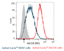
- Jurkat-Lucia™-hICOS: Reporter T cells
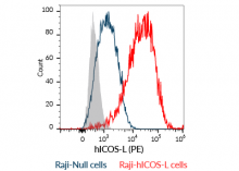
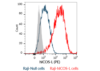
- Raji-hICOS-L: Antigen-presenting cells (APCs)
These paired cell lines allow the mimicking of the immune synapse between T cells and APCs through the interaction of cell surface molecules delivering T-cell activation signals.
Inducible Co-stimulator (ICOS, CD278) is an immunostimulatory IC and a member of the CD28 superfamily. Expression of ICOS is rapidly induced in CD4 and CD8 T cells upon their activation, whereas its ligand ICOS-L (CD275), is mostly expressed by APCs [1].
Assay principle:
This assay relies on the co-culture of Jurkat-Lucia™ hICOS and Raji-hICOS-L cells:
– Jurkat-Lucia™-ICOS cells express a hICOS-CD3ζ fusion protein along with the Lucia luciferase reporter gene under the control of an ISG54 minimal promoter fused to six NFAT response elements. CD3ζ is a key component of the T-cell receptor (TCR) and CD3 complex that triggers TCR downstream signaling.
– Raji-hICOS-L cells express the ICOS-L activating IC ligand.
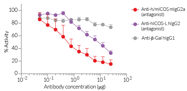
In the presence of a potent ICOS or ICOS-L antagonist, the ICOS-CD3ζ-mediated activation is blocked and there is no Lucia production. Activation of the reporter T cells can be readily measured using QUANTI-Luc™ 4 Lucia/Gaussia detection reagent (see Figures).
T-cell key features:
- Stable hICOS-CD3ζ expression
- NFAT-inducible Lucia luciferase reporter activity
APC key features:
- Stable hICOS-L overexpression
InvivoGen also offers Jurkat-Lucia™ hICOS cells without Raji-hICOS-L cells to measure the potency of agonists of the ICOS/ICOS-L axis.
![]() Read our review on Immune Checkpoint Blockade
Read our review on Immune Checkpoint Blockade
![]() Learn more about Immune Checkpoint Antibodies.
Learn more about Immune Checkpoint Antibodies.
Reference:
1. Amatore, F. et al. 2020. Role of ICOS in cancer immunotherapy. Expert Opin Biol Ther 20, 141-150.
Figures


Specifications
Growth medium: IMDM, 2 mM L-glutamine, 25 mM HEPES, 10% (v/v) heat-inactivated fetal bovine serum (FBS), 100 U/ml penicillin, 100 µg/ml streptomycin
Antibiotic resistance:
- Jurkat-Lucia™ hICOS cells: Blasticidin and Zeocin®
- Raji-hICOS-L cells: Blasticidin
Quality Control:
- Human ICOS and ICOS-L expression have been verified by flow-cytometry.
- Reporter activity has been validated following the co-culture of the two cell lines.
- The stability for 20 passages following thawing has been verified.
-
Both cell lines are guaranteed mycoplasma-free.
These products are covered by a Limited Use License (See Terms and Conditions).
Back to the topContents
Please note: Both cell lines are sold together.
- 3-7 x 10⁶ Jurkat-Lucia™ hICOS cells
- 3-7 x 10⁶ Raji-hICOS-L cells
- 1 ml of Blasticidin (10 mg/ml)
- 1 ml of Zeocin® (100 mg/ml)
- 1 tube of QUANTI-Luc™ 4 Reagent, a Lucia luciferase detection reagent (sufficient to prepare 25 ml)
![]() Shipped on dry ice (Europe, USA, Canada)
Shipped on dry ice (Europe, USA, Canada)
Details
Inducible Co-stimulator (ICOS, CD278) is an immunostimulatory IC and a member of the CD28 superfamily. Expression of ICOS is rapidly induced in CD4+ and CD8+ T cells upon their activation, whereas its ligand ICOS-L (also known as CD275), is mostly expressed by antigen-presenting cells [1]. The interaction between ICOS and ICOS-L delivers a secondary co-stimulatory signal through the activation of the transcription factor AKT, which promotes T cell proliferation and differentiation as well as the production of cytokines [1]. In tumor immunity, ICOS is involved in the amplification of the anti-tumor cytotoxic CD8+ T cell response, as well as the 'pro-tumor' function and maintenance of regulatory T cells (Tregs). Therefore, both agonistic and antagonistic monoclonal antibodies (mAbs) targeting this pathway are being investigated in combinational cancer immunotherapy [2]. Notably, ICOS agonistic mAbs have been shown to potentiate the effects of anti-CTLA-4 mAbs [3].
References:
1. Amatore, F. et al. 2020. Role of ICOS in cancer immunotherapy. Expert Opin Biol Ther 20, 141-150.
2. Solinas, C. et al. 2020. The rationale behind targeting the ICOS-ICOS ligand costimulatory pathway in cancer immunotherapy. ESMO Open 5.
3. Soldevilla, M.M. et al. 2019. ICOS Costimulation at the Tumor Site in Combination with CTLA-4 Blockade Therapy Elicits Strong Tumor Immunity. Mol Ther 27, 1878-1891.